Leg Bones Diagram / Bone Structure Anatomy And Physiology. These muscles work together to produce movements such as standing, walking, running, and jumping. The elbow is located below the chest at the back of the foreleg. This large tendon from the powerful thigh muscles (quadriceps) wraps round the patella and is attached to the top of the lower leg bone (tibia). The patella is the kneecap bone. The lower leg is comprised of two bones, the tibia and the smaller fibula.
2006 kia optima belt diagram. The lower leg is comprised of two bones, the tibia and the smaller fibula. Home » unlabelled » bones in leg diagram / your leg bones are very large and strong to help support the weight of your body. The major bones of the leg are the femur (thigh bone), tibia (shin bone), and adjacent fibula, and these are all long bones.the patella (kneecap) is the sesamoid bone in front of the knee.most of the leg skeleton has bony prominences and margins that can be palpated and some serve as anatomical landmarks that define the extent of the leg. The patella is the kneecap bone.
Patella Wikipedia from upload.wikimedia.org The hip itself is a ball and socket joint, much like the shoulder.the structures necessary to create this joint are the socket, the joint capsule, muscle, ligaments, and the neck. The elbow is located below the chest at the back of the foreleg. Anatomy of the foot (26/28 bones) 11 terms. The rounded, proximal end is the head of the femur, which articulates with the acetabulum of the hip bone to form the hip joint. Its lower end helps create the knee joint. These muscles work together to produce movements such as standing, walking, running, and jumping. Muscle anatomy in shoulder 12 photos of the muscle anatomy in shoulder muscle anatomy neck and shoulder, muscle anatomy of shoulder, muscle anatomy of shoulder joint, muscle anatomy shoulder back, muscle anatomy shoulder upper arm, human muscles, muscle anatomy neck and shoulder, muscle anatomy of shoulder, muscle. The lower leg extends from the knee to the ankle.
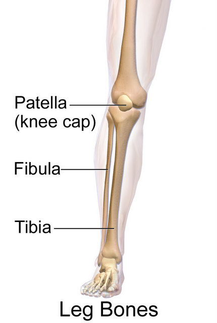
The bones of the leg are the femur, tibia, fibula and patella.the foot bones shown in this diagram are the talus, navicular, cuneiform, cuboid, metatarsals and calcaneus.
Joints of hand anterior view, lateral view, right hand. Its lower end helps create the knee joint. The tarsal bones in the foot are located amongst tibia, metatarsal bones, and fibula. The knee joint is the largest joint in the body and is primarily a hinge joint, although some sliding and rotation occur. These landmarks are the anterior superior iliac spine. The bones of the leg and foot form part of the appendicular skeleton that supports the many muscles of the lower limbs. This is the first joint in the leg. The bones of the leg are the femur, tibia, fibula and patella.the foot bones shown in this diagram are the talus, navicular, cuneiform, cuboid, metatarsals and calcaneus. Muscle system diagram photos muscular system more than 133 muscle chart back at on the anterior and posterior views of the muscular system above, superficial muscles (those at for the legs. These muscles work together to produce movements such as standing, walking, running, and jumping. The fibula is connected via ligaments. The diagram of bones in the ankle and foot is given below: 2006 kia optima belt diagram.
Then add shoulder blades, front legs. The knee joint is the largest joint in the body and is primarily a hinge joint, although some sliding and rotation occur. The elbow is located below the chest at the back of the foreleg. It lies within the quadriceps tendon. The tarsal bones in the foot are located amongst tibia, metatarsal bones, and fibula.
Broken Leg Tibia Fibula Settlement Amounts Car Accidents And More from www.justinziegler.net This is the first joint in the leg. Ulna and the radius are two bones that sit next to each other. At the same time, the bones and joints of the leg and foot must be strong enough to support the body's weight while remaining. The bones of the leg are the femur, tibia, fibula and patella.the foot bones shown in this diagram are the talus, navicular, cuneiform, cuboid, metatarsals and calcaneus. Then add shoulder blades, front legs. Home » unlabelled » bones in leg diagram / your leg bones are very large and strong to help support the weight of your body. The bones of the leg are the femur, tibia, fibula and patella.the foot bones shown in this diagram are the talus, navicular, cuneiform, cuboid, metatarsals and calcaneus. Its lower end helps create the knee joint.
The bones of the leg and foot form part of the appendicular skeleton that supports the many muscles of the lower limbs.
The bones of the leg are the femur, tibia, fibula and patella.the foot bones shown in this diagram are the talus, navicular, cuneiform, cuboid, metatarsals and calcaneus. The lower leg is comprised of two bones, the tibia and the smaller fibula. Joints of hand anterior view, lateral view, right hand. Then add shoulder blades, front legs. An update femur, tibia, fibula, calcaneous, tarsals, metatarsals, p make a model of the human skeleton anteriorleg skeleton arm clipart free download on clipartmag muscle blank drawing google search human anatomy and physiology, anatomy, anatomy and. Home » unlabelled » bones in leg diagram / your leg bones are very large and strong to help support the weight of your body. He leg's main function in the human is for use the leg bones diagrams to learn the names of the leg bones and leg anatomy. Its lower end helps create the knee joint. The rounded, proximal end is the head of the femur, which articulates with the acetabulum of the hip bone to form the hip joint. To explain the term in layman's language, it is the heel bone in the skeletal system. Disposition of rotator cuff muscles diagram. The bones of the leg and foot form part of the appendicular skeleton that supports the many muscles of the lower limbs. The bones together make up the hip.
Framework of bones, class 6. The lower leg is comprised of two bones, the tibia and the smaller fibula. Browse 7,052 leg bone stock photos and images available, or search for leg bone xray or human leg bone to find more great stock photos and pictures. The femur, or thighbone, is the longest and largest bone in the human body. The bones of the leg are the femur, tibia, fibula and patella.the foot bones shown in this diagram are the talus, navicular, cuneiform, cuboid, metatarsals and calcaneus.
Hip Thigh Atlas Of Anatomy from doctorlib.info These bones have a marrow, but not a bone marrow cavity. Find the gnaw marks on a bone where likely a rodent chewed. The femur, or thigh bone, is the single bone of the thigh region (figure 6.51). Bones of the lower limb · anatomy and physiology tri to cook: Framework of bones, class 6. The major bones of the leg are the femur (thigh bone), tibia (shin bone), and adjacent fibula, and these are all long bones.the patella (kneecap) is the sesamoid bone in front of the knee.most of the leg skeleton has bony prominences and margins that can be palpated and some serve as anatomical landmarks that define the extent of the leg. The back of the patella is covered with smooth cartilage. See more ideas about muscle anatomy, leg muscles anatomy, anatomy.
Your leg bones are very large and strong to help support the weight of your body.
It is made of the ulna and the radius. 2006 kia optima belt diagram. The lower leg extends from the knee to the ankle. The bones of the human leg, like those of other mammals, consist of a basal segment, the femur (thighbone); The patella is the kneecap bone. The femur, or thighbone, is the longest and largest bone in the human body. There are in all 7 bones, which fall under tarsal bones category. The bones of the hip include the femur, the ilium, the ischium, and the pubis. The forearm is the long bone that runs just after the elbow. This large tendon from the powerful thigh muscles (quadriceps) wraps round the patella and is attached to the top of the lower leg bone (tibia). The lower leg is comprised of two bones, the tibia and the smaller fibula. Your leg bones are very large and strong to help support the weight of your body. Disposition of rotator cuff muscles diagram.
Share :
Post a Comment
for "Leg Bones Diagram / Bone Structure Anatomy And Physiology"


Post a Comment for "Leg Bones Diagram / Bone Structure Anatomy And Physiology"